THE PLUS RHINOPLASTY IN KOREA
Implant Rhinoplasty in Korea

THE PLUS RHINOPLASTY IN KOREA
Implant Rhinoplasty in Korea
the plus rhinoplasty in Korea
Implant Rhinoplasty Surgery
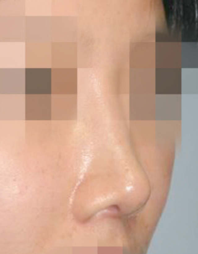
Implants are commonly used in East Asians with small noses. While using an implant is more convenient than using autologous (one’s own) tissue, it can also cause serious irreversible damage and deformity to the nose. Therefore, it is crucial to actively prevent complications from occurring, regardless of the type of implant used for nose surgery, and to provide appropriate treatment when they occur.
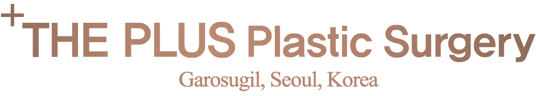
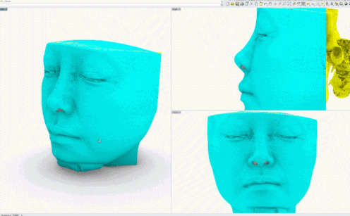
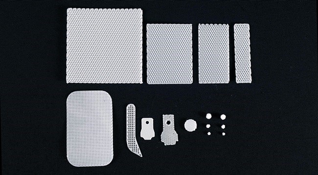
3D Custom Implants
The silicone implants commonly used in rhinoplasty are carved by the surgeon during the operation. However, with the recent advancement of 3D printing technology, custom implants are pre-made based on the patient’s CT images and used during surgery. This not only fits the patient’s nose exactly, but also reduces the time to carve the implant during surgery, which can shorten the operation time. The production period takes about a week, so after the CT scan at the consultation, the 3D implant production and surgery will proceed.
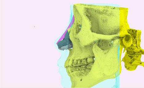
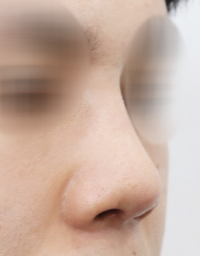
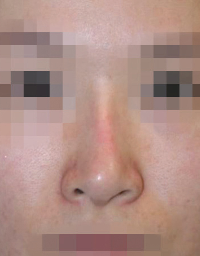
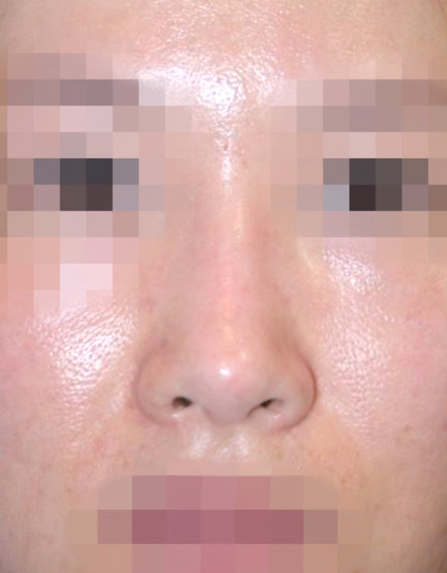
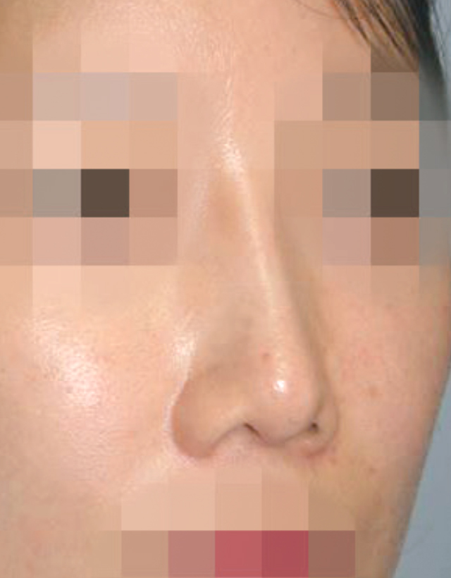
Before and After Cases of 3D Custom Implant Surgery
Artificial Implants
Case A: Silicone
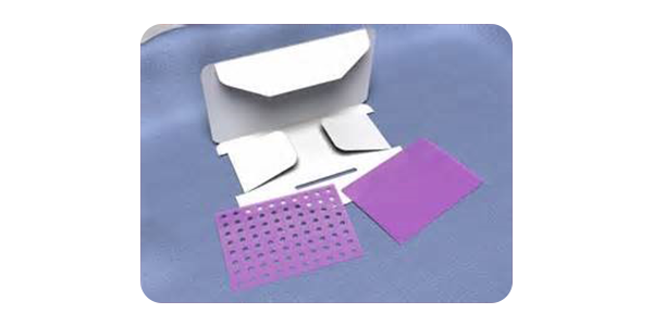
Silicone is the most long-standing and still the most widely used implant in the field of nose surgery. It has the advantage of being somewhat verified due to the development of the material itself and trial and error over a long period of time. In the past, solid silicone with high hardness was widely used, but recently, there is a trend to use solid silicone implants with low hardness and softness, like high soft silicone.
Advantage: The Plus Plastic Surgery also prefers silicone as an implant. Silicone is relatively easy to handle, allows delicate carving, and is mainly used because it is easy to remove and correct.
Disadvantage: Silicone implants are surrounded by a capsule composed of fibrous tissue after being transplanted into the body. This is a result of a normal immune response in the body, but this capsule sometimes acts as the core of severe deformation accompanied by contraction and inflammation. Also, there are cases where the implant escapes, gets infected, and if it is inserted for a long time, phenomena such as degeneration or calcification of the implant may occur.
Case B: Gore-Tex
In Korea, Gortex has been used in the field of nose surgery since the 2000s. It is an artificial implant that is often used in nose surgery along with the existing implant, silicone. Goretex is expanded polytetrafluoroethylene (ePTFE) with micropores, and after insertion, the surrounding tissue grows into the micropores, forming adhesion with the surrounding tissue.
Advantages: It has the advantage of less formation of surrounding membranes compared to silicone and no rejection reaction, and after insertion, various serum proteins in the blood attach to the surface and act as a mediator between inflammatory cells and fibroblasts, helping to adhere to surrounding tissues.
Disadvantages: There is a problem that if bacteria penetrate, it may not be able to defend properly. In fact, inflammation caused by Goretex has a greater ripple effect on surrounding normal tissues than inflammation of silicone implants, and inflammation control is not well done, so if inflammation of the Goretex implant occurs, it is better to remove it quickly.
Unnatural Cases of Goretex
See more before and after surgery photos.
Case C: Silitek or Chimera
The Silitek implant is a composite with the top made of Goretex and the bottom made of silicone, a product designed to maximize the advantages of the two types of implants. The top is closely attached to the skin with Goretex, and the bottom is positioned on the bone cartilage support structure with silicone. In other words, it shares the pros and cons of silicone and Goretex.
Pros: There is less chance of problems due to capsule formation on the skin side, and it adheres well to the tissue. The bottom side allows for fine carving.
Cons: In secondary surgery, it is easy for the junction of the two materials to separate and there may be situations where complete removal is not possible, so care is needed to ensure that no fragments are left when removing.
Correction after Silitek Removal
See more before and after surgery photos.
Case D: Medpor
Medpor is a material made of porous high density polyethylene and has been widely used in the field of plastic surgery for a long time, especially for facial reconstruction and especially bone tissue reconstruction. Because it is made of high density material, it has considerable strength. It has been emphasized that through these pores, surrounding tissues and new blood vessels grow in, forming fibrous tissue and stabilizing. Recently, as the frequency of nose reoperation increases, there are many cases of removing Medpor. In this case, Medpor is strongly adhered to the surrounding tissue, and it is common for the surrounding tissue including cartilage to harden, making it difficult to remove, and often needs to be supplemented with autologous cartilage after removal.
Case E: PCL Mesh (T&R Mesh)
Recently, PCL mesh (T&R Mesh) has been created based on 3D printing technology and is being used as a cartilage substitute. It has a melting property over several years and is a bit stronger than cartilage.
Before and After PCL Mesh
Click here to view details.
Case F: PDO Foil (PDS Plate)
PDO Foil is a recent addition to the materials used in nose surgery. It shares the same ingredients as the sutures used in the procedure. Its primary function is to supplement the strength of cartilage. It breaks down over several months and mimics the elasticity of cartilage.
Click here to view details.
Allograft (Donated Tissue From Another Person)
Currently, the allografts used in rhinoplasty include Alloderm, allograft costal cartilage, and allograft fascia lata. Despite their high cost, they are actively used in the field of rhinoplasty. They can be used in patients who have undergone multiple reoperations due to a lack of donor site. However, these patients have a higher incidence of absorption and infection, so they should be used appropriately depending on the situation.
Case A: Alloderm / Megaderm
Alloderm is an acellular dermal matrix made by selectively collecting only the dermis from the body skin and going through special immune treatment, for example freeze-drying. The basement membrane, elastin, and vascular channel, which are the original dermal structures, are preserved, so new blood vessels, nerves, collagen regeneration occur within the transplanted individual. This has the advantage of being replaced with autologous tissue in the long term.
Advantages: Alloderm, which is commonly used in nose surgery, is produced in various sizes and thicknesses, and it is very convenient to use as a graft for the nose bridge or nose tip alone or in conjunction with an implant. It can be very useful for rhinoplasty, nasal bridge grafting, thin skin supplementation, and prevention of implant or cartilage graft exposure.
Disadvantages: Irregular absorption can occur, so excessive thickness is not good. Therefore, it is not suitable for the role of raising a low nose bridge. It is mainly used to cover the skin around the nose tip or the nose tip graft so that it does not show.
Case B: Irradiated Homologous Costal Cartilage (IHCC)
Irradiated Homologous Costal Cartilage is made safe from infectious diseases by destroying all cells, bacteria, and viruses through the manufacturing process of cell membrane destruction. This can be achieved through osmotic pressure, protein denaturation using hydrogen peroxide or alcohol, radiation exposure, or using chest cartilage collected from donated bodies. The role of a strong support for the correction of a short nose for Asians or a collapsed nose is taken by the septal cartilage or chest cartilage, but recently, Irradiated Homologous Costal Cartilage is being used as an alternative for patients who have difficulty collecting cartilage.
Advantages:
• Surgery time is shortened.
• Recovery is faster and patient pain is reduced because cartilage collection is not necessary.
• Additional chest scars can be reduced.
Disadvantages:
• The absorption rate is unclear (the deviation of the absorption rate varies depending on the condition of the donor).
• Since it is not autologous (one’s own) tissue, side effects such as inflammation and infection can be problematic
• If used on the tip of the nose, tip hardness occurs.
Case C: Homologous Fascia Lata
Homologous fascia lata is a product that has been commercialized by treating antigens using osmotic pressure, drug treatment, and radiation on the fascia lata collected from a corpse. It can be used to supplement the bridge of the nose or irregular parts of the skin. It is not commonly used.
Xenograft (Donated Animal Tissue)
So far, the supplied xenograft products have the disadvantage of being expensive and inconvenient to purchase, and are not often used in common primary or secondary surgeries. In addition, patient’s can be opposed to using tissues extracted from animals. As the supply and use of homografts increase, xenografts are not commonly used these days.
Case A: Lyoplant
Lyoplant is made by extracting collagen through mechanical and chemical treatment, for example freeze-drying, of the a calf’s pericardium. The connective tissue is loosely maintained so that it can easily fuse with the human body. It is very thin and has a high absorption rate, so it is recommended to use it restrictively.
Click here to view details.
Case B: Permacol
Permacol is an acellular dermal matrix product that removes antigens from pig dermis. From the author’s experience, compared to autologous tissue or homografts such as alloderm, it was observed that scar formation with surrounding tissues was severe and the tissue became hard. It is very vulnerable to inflammation, so if inflammation is suspected after transplantation, it is recommended to remove it immediately.
Click here to view details.
View the research papers from The Plus Plastic Surgery medical team.
Frequently Asked Questions
Why is silicone commonly used for rhinoplasty at THE PLUS PS in Gangnam, South Korea?
Silicone is commonly used at THE PLUS PS because it is easy to handle, allows for delicate carving, and is known for its long-standing reliability. The clinic prefers silicone implants as they are easy to remove and correct if necessary, making them a popular choice for rhinoplasty procedures.
What are the benefits of using 3D printed custom implants for nose surgery?
3D printed custom implants are tailored to fit the patient’s nose precisely, based on their CT images. This advancement not only reduces the time needed to carve the implant during surgery, potentially shortening operation time, but also improves the overall accuracy and fit of the implant. The custom implant production typically takes about a week after a CT scan is performed.
What precautions are taken to prevent complications from nose implants at THE PLUS PS?
THE PLUS PS emphasizes the importance of preventing complications by selecting the appropriate type of implant and ensuring meticulous surgical techniques. In the event of any complications, the clinic is prepared to provide proper and timely treatment to address issues such as implant contraction or inflammation. The choice of softer silicone implants also helps minimize potential deformation and immune reactions.